Mitotic Spindle and Cell Cycle Rate My Science Biology Diagrams (A) Features of the metaphase mitotic spindle. With their minus ends tethered at the spindle poles, microtubules extend either to the kinetochores of paired chromatids (kinetochore fibers), to the central spindle where they form an overlapping antiparallel array (interpolar microtubules), or away from the spindle towards the cell cortex (astral microtubules). The mitotic spindle in yeast (A, left) is formed from spindle pole bodies (A, right) that are composed of five subcomplexes (B). (A, left) Immunofluoresence of a large-budded mitotic yeast cell showing SPBs marked by Spc42-GFP (green), microtubules (red), and DNA (blue) and electron micrograph (A, right) showing trilaminar ultrastructure.
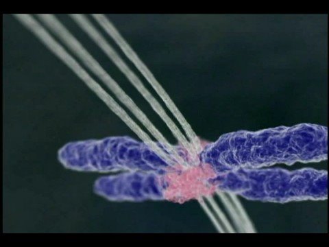
In all eukaryotes, morphogenesis of the microtubule cytoskeleton into a bipolar spindle is required for the faithful transmission of the genome to the two daughter cells during division. This process is facilitated by the intrinsic polarity and dynamic properties of microtubules and involves many proteins that modulate microtubule organization and stability. Recent work has begun to uncover
Mitotic spindle assembly in animal cells: a fine balancing act Biology Diagrams
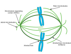
The assembly and dynamics of the mitotic spindle rely on the shifting balance between opposing plus-end-directed and minus-end-directed motor proteins. Three classes of spindle microtubules can be distinguished in mitotic animal cells they play an active part in spindle formation. The influence of the chromosomes can be demonstrated by Mitotic spindle formation begins as cells transition from interphase into mitosis. As the nuclear envelope breaks down in prophase, microtubules emanating from centrosomes undergo rapid polymerization and depolymerization, probing the intracellular space for chromosomes. This dynamic behavior, driven by tubulin instability, enables efficient

Mitotic spindle formation is a critical event that takes place during prophase. Several events of mitosis depend on the mitotic spindle, which forms in the cytoplasm during prophase.

Definition, Formation & Function - Study.com Biology Diagrams
The mitotic spindle is a microtubule-based assembly that separates the chromosomes during cell division. The life of a spindle starts with its formation by interactions between microtubules and chromosomes mediated by the microtubule-associated proteins and the kinetochore proteins during prometaphase and ends with chromosome segregation in A computational model for the formation of lamin-B mitotic spindle envelope and matrix. Interface Focus 4 , 20130063 (2014). Article PubMed PubMed Central Google Scholar
