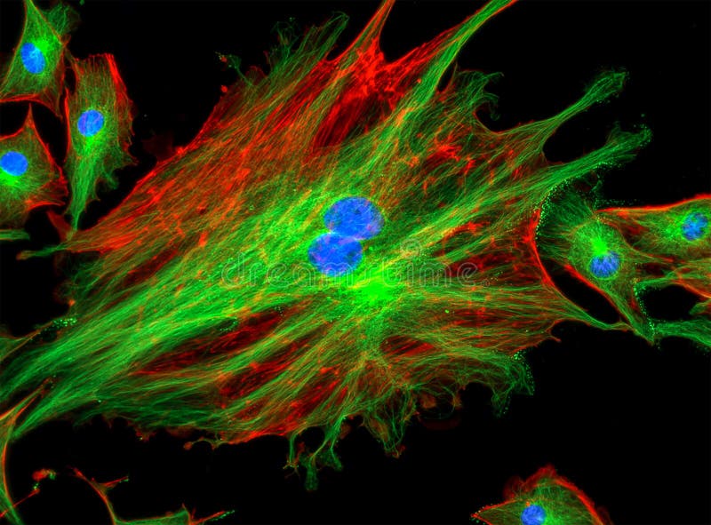
mitosis cells stained with fluorescent dyes Biology Diagrams 1.1. OVERVIEW OF FLUORESCENCE IMAGING AND MITOSIS. In the late 1800's Flemming, Van Beneden and their contemporaries carried out the first studies of chromosome behavior in dividing cells in both living and fixed tissue using the available microscope methodology . Since that time tremendous advances in microscope technology and fluorescence 1.1 Overview of Fluorescence Imaging And Mitosis. In the late 1800's Flemming, Van Beneden and their contemporaries carried out the first studies of chromosome behavior in dividing cells in both living and fixed tissue using the available microscope methodology (reviewed in ). Since that time, tremendous advances in microscope technology and The impact of fluorescence microscopy on cell biology in general, and on the field of mitosis in particular, is profound. This major impact has occurred due to the confluence of the development of fluorescent reporter molecules that can be delivered to and expressed in live cells and advances in equipment used for imaging.

Fluorescence microscopy is one of the most important approaches in the cell biologist's toolbox for studying the mitotic spindle. In fact, many of the key insights into our understanding of mitosis have been enabled by the visualization of mitotic processes using fluorescence microscopy. Here, we su … Mitosis is a highly dynamic and choreographed process in which chromosomes are captured by the mitotic spindle and physically segregated into the two daughter cells to ensure faithful transmission of the genetic material. Live-cell fluorescence microscopy enables these dynamics to be analyzed over diverse temporal scales.

Live fluorescence imaging of chromosome segregation in cultured cells Biology Diagrams
Both LSCM and SDCM systems are available with multiple lasers for imaging a range of fluorescent probes. A third alternative for imaging mitosis is to use a wide-field fluorescence microscope and perform deconvolution to remove out-of-focus fluorescence . Turnkey systems are available for this mode of microscopy (e.g. DeltaVision). Mitosis is a highly dynamic and choreographed process in which chromosomes are captured by the mitotic spindle and physically segregated into the two daughter cells to ensure faithful transmission of the genetic material. Live-cell fluorescence microscopy enables these dynamics to be analyzed over diverse temporal scales. 73.0K Views. Mitosis is a form of cell division in which a cell's genetic material is divided equally between two daughter cells. Mitosis can be broken down into six phases, during each of which the cell's components, such as its chromosomes, show visually distinct characteristics. Advances in fluorescence live cell imaging have allowed scientists to study this process in great detail